

Could Breast Tissue Stiffness Help Explain the Increased Cancer Risk in Women with Dense Breasts?
Women with dense breast tissue face up to a four-fold higher lifetime risk of developing breast cancer. Dense breasts feel firm because they contain a tight mesh of structural collagen proteins, stiffer tissue architecture, and an increased number of immune cells called macrophages. This structural firmness and tissue stiffness are well-known physical features of aggressive breast tumors and may be linked to treatment resistance and cancer spread.
While doctors have long recognized that dense tissue and chronic inflammation are linked to higher cancer risk, the exact physical process connecting tissue firmness to genetic DNA damage is not fully understood. Previous research has shown that macrophages can produce reactive oxygen species (ROS) – molecules that can damage cells – but scientists did not know how stiff breast tissue might change the behavior of macrophages in a way that ultimately damages the DNA of nearby breast epithelial cells.
This study, led by researchers at UCSF, uncovers a clear, step-by-step chain reaction showing how physical tension in firm tissue causes breast cells to recruit immune cells (macrophages), which then produce toxic chemical byproducts that damage DNA and thereby drive tumor development.
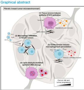
Image source: Tissue tension fosters macrophage-driven lipid peroxidation-induced DNA damage
Study Description
This research combined human patient data, tissue samples, and specialized animal and cell models. The researchers analyzed genetic data from human breast cancer databases (TCGA-BRCA) and examined tissue samples removed during prophylactic mastectomy from women with high or low mammographic density. They also studied breast cancer in mice using various genetic modifications—including removing stress-response proteins in breast cells, deleting protective antioxidant enzymes in immune cells, and using specialized antibodies to neutralize altered fats. Advanced tools like Atomic Force Microscopy (AFM) and machine-learning mapping (STIFMap) were used to measure microscopic tissue firmness.
Key Findings
- Stiffer breast tumors were associated with more genetic changes and mutations.¹
- When breast tissue becomes stiff and fibrotic, it creates physical tension on surrounding cells. This mechanical pull activates a key stress-response protein inside breast cells called STAT3. Activated STAT3 causes breast cells to release chemical signals (chemokines like CCL3 and CSF1) that act like beacon flares, attracting immune defender cells called macrophages into the dense tissue.2
- As these macrophages squeeze into the stiff environment, physical pressure forces them to produce high levels of unstable oxygen molecules (reactive oxygen species, or ROS) via an enzyme called NOX.3
- These oxygen molecules react with fats in nearby cell membranes (a process called lipid peroxidation). This fat breakdown creates small, highly reactive chemical compounds called lipid aldehydes (such as HNE and acrolein). Unlike short-lived oxygen molecules, these toxic aldehydes easily drift into neighboring breast cells and enter the nucleus, binding to DNA and causing double-strand breaks and genetic mutations.4
- Blocking these oxidized fats or lipid aldehydes reduced DNA damage and tumor growth in experimental models.⁵
- Breast tissue from women with high mammographic density had more macrophages, more evidence of lipid aldehydes, and more DNA damage than tissue from women with low mammographic density.⁶
By demonstrating that tissue firmness activates a stress signal that draws in macrophages to create toxic fat byproducts, this study connects physical tissue changes directly to DNA mutations. It reveals that dense breast tissue is not just an inactive background feature, but an active player in generating genetic damage. Clinically, this discovery opens up exciting new possibilities for prevention. While it is difficult to physically soften dense tissue, these results show that we can target the chemical steps in the middle—such as using treatments that absorb toxic fat byproducts or neutralize oxidized lipids—to protect DNA and lower cancer risk in women with dense breasts.
Limitations
Much of the evidence showing how tissue stiffness leads to DNA damage came from breast cancer cells and mouse models. Although these experiments help researchers understand the individual steps involved, results from laboratory and animal models do not always translate directly to people. The study shows that dense breast tissue had more macrophages, lipid aldehydes, and DNA damage, but it cannot prove that breast density or tissue stiffness caused those changes or that women with these findings would later develop breast cancer. Larger and longer-term studies in women are needed to confirm the pathway and determine its importance in breast cancer development.
Detailed Results
¹ Analysis of human breast cancers from The Cancer Genome Atlas (TCGA) found that greater tumor fibrosis was significantly associated with higher tumor mutational burden after accounting for breast cancer subtype, TP53 mutation status, and tumor purity (P=.04). In mouse models, stiffer breast tumors also showed a broader range of genetic changes affecting protein-coding genes.
² Greater tissue stiffness increased activation of STAT3, a protein involved in cell signaling, and increased signals that attract macrophages. When STAT3 was removed from mammary epithelial cells in mice, macrophages in the surrounding tissue decreased from approximately to 20% (P<.001), and breast epithelial cells showing DNA damage decreased from approximately 34% to 18% (P<.01).
³ Macrophages grown on stiff surfaces (6 kPa) produced significantly more ROS than those grown on soft surfaces (400 Pa). In stiff mouse breast tumors, oxidation of fats within macrophages was approximately 1.5 times higher than in softer tumors (P<.001).
⁴ Exposing breast cancer cells to the lipid aldehyde 4-hydroxy-nonenal (HNE) produced increasing DNA damage with increasing exposure. L-carnosine, which can bind lipid aldehydes, reduced this damage (P<.0001). Human breast tumors with greater fibrosis also showed more evidence of mutations associated with exposure to the lipid aldehyde acrolein (P<.001).
⁵ In mice, neutralizing oxidized fats reduced the percentage of breast epithelial cells showing DNA damage from approximately 25% to 11% (P<.01) and reduced the relative area occupied by tumor from approximately 28% to 14% (P<.001). Conversely, genetically increasing lipid oxidation in macrophages increased DNA damage and lung metastases.
⁶ In prophylactic mastectomy specimens, breast tissue from women with high mammographic density had significantly more CD68+ and CD163+ macrophages than tissue from women with low density (P<.001). High-density tissue also had more epithelial cells containing HNE, a marker of lipid aldehyde exposure (P<.05), and more epithelial cells showing DNA damage (P<.001).