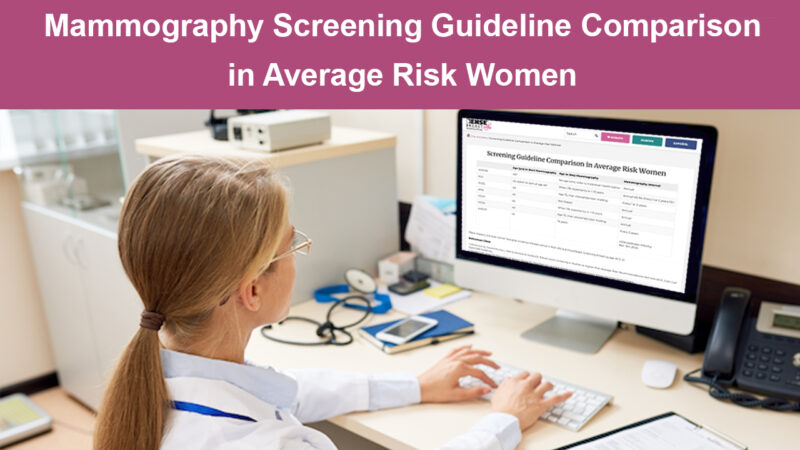
Mammo Screening Guideline Comparison Table, Silver Medal of Honor, New Studies
Comparison In Average Risk Women
This popular DBI table just updated to include the new ACOG recommendation that mammograms begin at age 40. Visit the table for comparative mammography recommendations by organization on recommended age to start, stop, and frequency.
Congratulations!
On October 24, 2024, Dr. Wendie Berg, Chief Scientific Advisor to DenseBreast-info.org, received the prestigious Silver Medal of Honor from the Austrian Medical Society to recognize her contributions to the design of the Austrian Breast Cancer Screening Program (which includes screening ultrasound for women with dense breasts).
New Studies:
-Breast Density and Sensitivity of Digital Mammography
Consistent with many prior analyses, Payne et al reported in a single-center UK study that digital mammography sensitivity decreases with increasing breast density as assessed by Volpara software, at 75% for grade a, 74% for grade b, 60% for grade c, and 51% for grade d. Symptomatic interval cancer rates at three years increased from 1.8 per 1000 for grade a to 3.2, 5.7, and 7.9 per 1000 for density grades b, c, and d, respectively.
-CEM for Breast Screening
A retrospective, single-institution study by Sorin et al. from Tel-Aviv University evaluated performance of CEM for breast cancer screening over a 10-year period in 5,424 women at intermediate or high risk for breast cancer (15-20% lifetime risk and >20% lifetime risk, respectively), with the following results:
- Most women (85%) included had dense breasts
- Contrast enhancement increased cancer detection compared to the low-energy images (which are similar to a standard 2D mammogram)1,2
- Specificity for CEM increased from the first to the third screening round, likely due to familiarity with the modality and availability of prior exams for comparison3
This study supports the viability of CEM as an effective screening modality for women with dense breasts and/or at intermediate breast cancer risk, and for high-risk women for whom MRI is not possible.
1Sensitivity for contrast-enhanced CEM images was 96% (71/74) compared to 58% (43/74) for low-energy images alone.
2The added cancer detection rate for CEM compared to the low energy images was 5.2 per 1000 screens (13.1/1000 for CEM vs. 7.9/1000 for the low energy images).
3Specificity overall was 81.8% (4378/5350); patient-level analysis showed improvement from 79.2% (2717/3431) in the first round to 89.2% (412/462) in subsequent rounds.

