
| QUICK FACTS |
Artificial Intelligence (AI) has many potential applications in breast cancer screening:
|
Quantifying Breast Density
Assessment of breast density can vary between radiologists, especially when the density is near the borderline between scattered fibroglandular (which is “not dense”) and heterogeneously dense (which is “dense”)[1]. Changes in reported breast density between dense and not-dense categories may affect eligibility and recommendations for supplemental screening in addition to mammography. AI can accurately and reproducibly quantify breast density [2–5] and provide more consistent results than radiologists [6].
Breast density assessment software adds costs to facilities and is not separately reimbursed by insurance carriers, nor are radiologists required to use it. Radiologist’s visual density assessment and AI software approaches show similar performance in identifying risk of screen-detected and interval cancer [7,8]. There are multiple commercial AI tools that are FDA approved for density assessment, and also some that are freely available (see Table 1).
Cancer Detection
AI improves cancer detection for both breast imaging specialists and general radiologists [9,10] by an average of 1.0-1.4 cancers per 1000 mammograms [11,12]. Importantly, AI does not generally increase the number of false positive examinations [9,11–15].
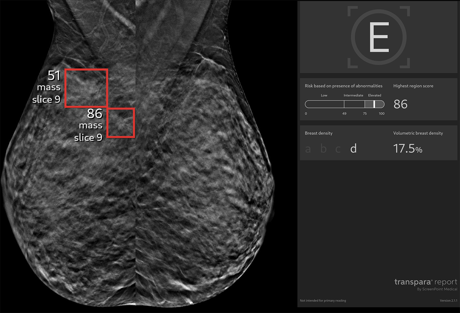
Breast density does not adversely affect cancer detection by AI (Figure 1). Greater improvement in cancer detection has been seen when AI is used in women with dense breasts than in women with non-dense breasts [9,16,17]. AI is successful in identifying early breast cancer. In one study, AI identified 16 of 21 (76%) T1 cancers, 20 mm or smaller, that had been missed by radiologists [9]. AI improves the detection of node-negative invasive cancers compared with radiologists alone [9].
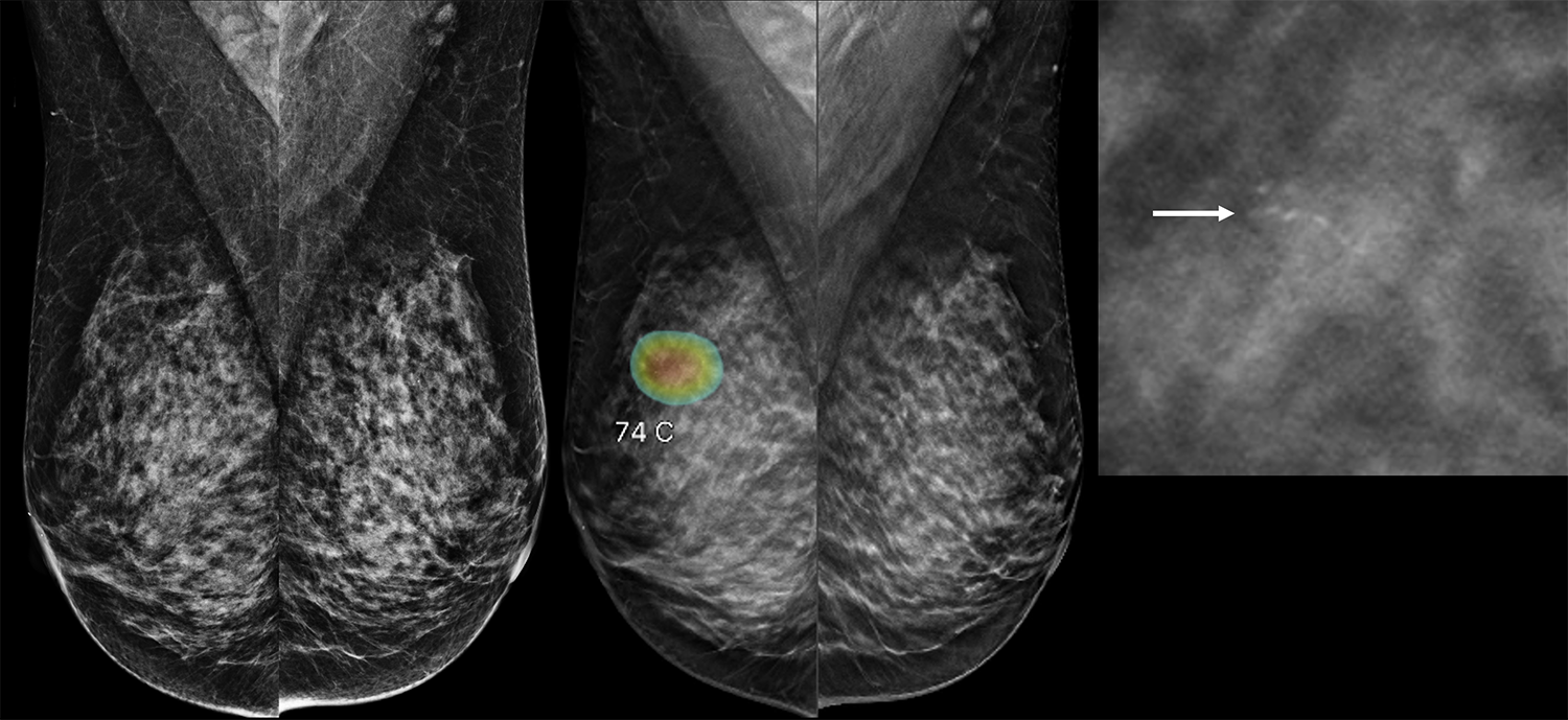
AI software varies and evolves but generally demonstrates high detection performance for the HER2-enriched subtype, conspicuous masses with microcalcifications [18,19] (Figure 2), and architectural distortion, with the ability to flag subtle abnormalities two to four years prior to clinical diagnosis [20]. However, AI is more likely to miss luminal and triple-negative subtypes, particularly when associated with favorable prognostic factors like smaller tumor size (≤ 2 cm) and lower histologic grade or relatively benign imaging features such as masses with circumscribed margins [19]. The primary reasons for missed cancers by AI include lesions obscured by overlapping dense breast tissue, located outside the mammary zone, or presenting with subtle mammographic features such as asymmetry.
Interval Cancers
Interval cancers are those found because of symptoms in the interval after a normal screening mammogram and before the next screen (one year in the United States). Interval cancers are much more common in women with dense breasts than in women with fatty breasts [21–23]. Some interval cancers were present but not seen on the prior mammogram and some newly develop. Interval cancers are often more aggressive and have worse outcomes than cancers found on a screening mammogram [24,25]. Retrospective studies show that on prior mammograms that had originally been interpreted as negative by radiologists, AI can identify 27% to 58% of subsequent symptomatic interval cancers [11,26–29]. One prospective randomized trial in Sweden showed fewer interval cancers with AI support than without, and interval cancers were less likely invasive or non-luminal than in the control group, but differences were not significant [30].
Costs
AI cancer detection software adds costs to facilities and is not separately reimbursed by insurance carriers, nor are radiologists required to use it. Some facilities in the United States offer AI review of the mammogram for an added charge, and others are integrating it into their practice without additional charge. Standalone AI has been shown to have performance similar to breast imaging specialists, but for legal and ethical reasons, as well as patient preference [31], the final interpretation of the mammogram is always done by a radiologist.
Improved Efficiency
AI can be used to prioritize (or “triage”) the reading order of mammograms based on likelihood of cancer, and it has been shown to reduce interpretation time [32,33]. In Europe, where many countries have required two radiologists to review all mammograms [34,35], AI is now replacing the second reader, and overall performance is slightly improved [12,14,32,36,37]. AI noticeably reduces screening workload and helps address workforce shortages [12,14,34,38,39].
Limitations
Some AI software has only been validated on 2D mammograms and not yet on 3D (tomosynthesis) mammograms (see Table 1). Comparisons between commercially available AI software have been made and there are substantial variations in performance, but algorithms continue to evolve. At present, only some of the AI tools make comparison to prior mammograms. When available, AI comparison to priors is limited to suspicious findings identified on the current mammogram (and that comparison can affect the AI “score”). Relatively benign-appearing cancers that humans identify because they are new are mostly missed by AI. Finally, the performance of AI software in women with prior breast-conserving treatment for breast cancer is poor [40].
Risk Assessment
Historically, the American Cancer Society [41], American College of Radiology, and National Comprehensive Cancer Network (NCCN) have defined “high risk” as an estimated lifetime risk of breast cancer of ≥20-25% (where average risk is about 12.5%). Lifetime risk is estimated using risk models such as Tyrer-Cuzick (IBIS). Such risk assessment is used to determine which women are recommended to have supplemental screening MRI and should not be used to determine when to start screening. Lifetime risk decreases as women age. About 20% of women in their 40s and 5% of women in their 60s are considered “high risk” by this definition, with recommendation for annual screening MRI in addition to mammography. It is uncommon for women over age 70 to be considered high risk.
AI can be used as an alternative to risk models. By reviewing the mammographic images alone, without clinical or demographic risk factors such as family history or prior biopsy history, AI software can be used to estimate the short-term risk of developing breast cancer in the next 1, 2, 5, or even 10 years [5,42]. These algorithms quantify breast density and “complexity” [43,44] as well as subtle imaging patterns and tissue characteristics that are not discernible to human readers. Explicitly adding breast density, family history, and other risk factors generally does not improve performance beyond AI alone [42,45]. An increase in AI risk score over time increases the likelihood cancer is present [46].
AI risk prediction is slightly more accurate than standard risk models [45]. Current NCCN guidelines state that the Gail model or AI image-based risk assessment can be used to identify women with a 5-year risk of breast cancer of ≥ 1.7% and that such women can “consider” supplemental screening with MRI (see “limitations” of this approach below). Adding polygenic risk score dramatically improves Gail model performance and may modestly improve AI performance [47].
MRI access is limited, and the alternative of contrast-enhanced mammography is not yet FDA approved for screening. AI could help by identifying women at higher risk who would benefit most from screening MRI. This could make use of MRI more efficient.
In the ScreenTrustMR study in Sweden, the risk scores from three models were averaged and the top 6.9% of women were randomized to invitation to MRI screening. Of 663 women invited, 559 (84%) had MRI, with a cancer detection rate of 64/1000 examinations [48]. This cancer detection rate is greatly enriched compared to the DENSE trial yield from MRI of 16.5/1000 [49]. Across multiple series [2–5,16,50,51], if MRI is recommended only in the top tier of risk (e.g. the top 4-12%), then from 20-59% of women with cancer will receive MRI (see Table 2). These tools are more effective than density alone at identifying women at risk who would benefit from supplemental MRI, and it is easier to implement AI review than manual risk model calculations.
Limitations: At best, only about 70% of women who will develop breast cancer in the ensuing 5 years will be identified by use of either existing risk models or AI software. Restricting MRI screening recommendations to those women at high risk based on AI alone would exclude some women with dense breasts and cancer that might be hidden on mammography. Most software has been validated only on 2D mammograms, and not 3D mammograms (tomosynthesis) [42]. There is variability in performance of AI risk tools and confusion about implementation. No matter what method is used to estimate risk, MRI capacity remains limiting and is particularly less available in rural areas. Many AI tools are not yet FDA approved (see Table 2) and performance across diverse races and ethnicities is often not reported. Patients can send their mammograms to FDA-approved Clairity, and, for a fee, receive their estimated 5-year risk, but facilities may not yet have a consistent policy for using this information to recommend MRI, and appropriate thresholds remain a topic of discussion (see below).
Most AI risk tools are designed and validated to provide a 1- to 5-year risk estimate, though newer research [5] is addressing 10-year risk prediction; the optimal timeframe to guide supplemental screening recommendations is evolving. The appropriate threshold for five-year risk is also a matter of debate. Because 5-year risk increases with age, nearly all women over age 58 could meet the NCCN criterion of ≥1.7% 5-year risk, even though mammography alone is adequate for most women (especially if the breasts are not dense). Colditz et al [52] and the USPSTF suggest that an average 5-year risk ≥ 3.16% (or ≥ 3% per USPSTF) better aligns with 20% lifetime risk threshold for purposes of supplemental screening. Even so, in younger women, the 5-year risk cut-point corresponding to a 20% lifetime risk would be lower, at 1.34% for women age 40-44, 2.05% for women 45-49, and 2.6% for women 50-54 years of age. For older women, it would be higher, at 3.3% for women aged 55-59, 4.7% for women 60-64, 6.9% for women 65-69 and as high as 10.9% for women 70-74 [52].
1. Sprague BL, Conant EF, Onega T. Variation in Mammographic Breast Density Assessments Among Radiologists in Clinical Practice: A Multicenter Observational Study. Ann Intern Med. 2016;165:457-464.
2. Yala A, Mikhael PG, Strand F. Multi-Institutional Validation of a Mammography-Based Breast Cancer Risk Model. J Clin Oncol. 2022;40(16):1732-1740. doi:10.1200/JCO.21.01337
3. Yala A, Lehman C, Schuster T. A Deep Learning Mammography-based Model for Improved Breast Cancer Risk Prediction. Radiology. 2019;292(1):60-66. doi:10.1148/radiol.2019182716
4. Eriksson M, Destounis S, Czene K. A risk model for digital breast tomosynthesis to predict breast cancer and guide clinical care. Sci Transl Med. 2022;14(644). doi:10.1126/scitranslmed.abn3971
5. Eriksson M, Czene K, Scott C. A long-term image-derived AI-based risk model for primary prevention of breast cancer in individuals at high risk. Sci Transl Med. 2026;18(850). doi:10.1126/scitranslmed.ady7414
6. Holley SO, Cardoza D, Matthews TP, et al. Artificial intelligence and consistency in patient care: a large-scale longitudinal study of mammographic density assessment. BJR Artif Intell. 2025;2(1):ubaf004. doi:10.1093/bjrai/ubaf004
7. Destounis S, Johnston L, Highnam R, Arieno A, Morgan R, Chan A. Using volumetric breast density to quantify the potential masking risk of mammographic density.AJR. Am J Roentgenol2017. 208:222-227.
8. Kerlikowske K, Scott CG, Mahmoudzadeh AP. Automated and clinical breast imaging reporting and data system density measures predict risk for screen-detected and interval cancers: A case-control study.Ann Intern Med2018. 168:757-765.
9. Kim HE, Kim HH, Han BK. Changes in cancer detection and false-positive recall in mammography using artificial intelligence: a retrospective, multireader study. Lancet Digit Health. 2020;2(3). doi:10.1016/S2589-7500(20)30003-0
10. Lee SE, Han K, Yoon JH, Youk JH, Kim EK. Depiction of breast cancers on digital mammograms by artificial intelligence-based computer-assisted diagnosis according to cancer characteristics. Eur Radiol. 2022;32(11):7400-7408. doi:10.1007/s00330-022-08718-2
11. Lång K, Josefsson V, Larsson AM, et al. Artificial intelligence-supported screen reading versus standard double reading in the Mammography Screening with Artificial Intelligence trial (MASAI): a clinical safety analysis of a randomised, controlled, non-inferiority, single-blinded, screening accuracy study. Lancet Oncol. 2023;24(8):936-944. doi:10.1016/S1470-2045(23)00298-X
12. Hernström V, Josefsson V, Sartor H. Screening performance and characteristics of breast cancer detected in the Mammography Screening with Artificial Intelligence trial (MASAI): a randomised, controlled, parallel-group, non-inferiority, single-blinded, screening accuracy study. Lancet Digit Health. 2025;7(3). doi:10.1016/S2589-7500(24)00267-X
13. Nepute JA, Peratikos M, Toledano AY. Improved Breast Cancer Detection with Artificial Intelligence in a Real-World Digital Breast Tomosynthesis Screening Program. Clin Breast Cancer. Published online 2025. doi:10.1016/j.clbc.2025.05.007
14. Dembrower K, Crippa A, Colón E. Artificial intelligence for breast cancer detection in screening mammography in Sweden: a prospective, population-based, paired-reader, non-inferiority study. Lancet Digit Health. 2023;5(10). doi:10.1016/S2589-7500(23)00153-X
15. Chang YW, Ryu JK, An JK. Artificial intelligence for breast cancer screening in mammography (AI-STREAM): preliminary analysis of a prospective multicenter cohort study. Nat Commun. 2025;16(2248). doi:10.1038/s41467-025-57469-3
16. Liu Y, Sorkhei M, Dembrower K. Use of an AI score combining cancer signs, masking, and risk to select patients for supplemental breast cancer screening. Radiology. 2024;311(1). doi:10.1148/radiol.232535
17. Koch HW, Larsen M, Bartsch H, Kurz KD, Hofvind S. Artificial intelligence in BreastScreen Norway: a retrospective analysis of a cancer-enriched sample including 1254 breast cancer cases. Eur Radiol. 2023;33(5):3735-3743. doi:10.1007/s00330-023-09461-y
18. Chang YW, Ryu JK, An JK, Choi N, Park YM, Ko KH. Breast Cancers Detected and Missed by AI-CAD: Results from the AI-STREAM Trial. Radiol Artif Intell. 2026;8(1):e250281. doi:10.1148/ryai.250281
19. Woo OH, Song SE, Choe SJ, Kim M, Cho KR, Seo BK. Invasive Breast Cancers Missed by AI Screening of Mammograms. Radiology. 2025;315(3):e242408. doi:10.1148/radiol.242408
20. Martiniussen MA, Bergan MB, Kristiansen MU, et al. Location of AI risk markers and associated mammographic features in screening mammograms obtained years before screen-detected breast cancer. Eur Radiol. Published online June 18, 2026. doi:10.1007/s00330-026-12689-z
21. Oluyemi ET, Grimm LJ, Goldman L, et al. False-Negative Screening and Diagnostic Mammograms in the National Mammography Database From 2010 to 2022. AJR Am J Roentgenol. 2026;226(2):e2533636. doi:10.2214/AJR.25.33636
22. Boyd NF, Guo H, Martin LJ, et al. Mammographic density and the risk and detection of breast cancer. N Engl J Med. 2007;356(3):227-236. doi:10.1056/NEJMoa062790
23. Ciatto S, Visioli C, Paci E, Zappa M. Breast density as a determinant of interval cancer at mammographic screening. Br J Cancer. 2004;90(2):393-396. doi:10.1038/sj.bjc.6601548
24. MacInnes EG, Duffy SW, Simpson JA, et al. Radiological audit of interval breast cancers: Estimation of tumour growth rates. Breast. 2020;51:114-119. doi:10.1016/j.breast.2020.03.006
25. Niraula S, Biswanger N, Hu P, Lambert P, Decker K. Incidence, Characteristics, and Outcomes of Interval Breast Cancers Compared With Screening-Detected Breast Cancers. JAMA Netw Open. 2020;3(9):e2018179. doi:10.1001/jamanetworkopen.2020.18179
26. Bahl M, Langarica S, Lamb LR. Commercial Artificial Intelligence (AI) Tool for Screening Digital Breast Tomosynthesis: Factors Associated With AI-Based Breast Cancer Detection. AJR Am J Roentgenol. 2026;226:e2533792. doi:10.2214/AJR.25.33792
27. Gialias P, Wiberg MK, Brehl AK. The use of artificial intelligence (AI) to safely reduce the workload of breast cancer screening: a retrospective simulation study. Acta Radiol. Published online 2025. doi:10.1177/02841851251356176
28. Seker ME, Koyluoglu YO, Ozaydin AN. Diagnostic capabilities of artificial intelligence as an additional reader in a breast cancer screening program. Eur Radiol. 2024;34(9):6145-6157. doi:10.1007/s00330-024-10661-3
29. Bahl M, Langarica S, Lamb LR, Kniss AS, Do S. AI to Reduce the Interval Cancer Rate of Screening Digital Breast Tomosynthesis. Radiology. 2025;316(1):e241050. doi:10.1148/radiol.241050
30. Gommers J, Hernström V, Josefsson V, et al. Interval cancer, sensitivity, and specificity comparing AI-supported mammography screening with standard double reading without AI in the MASAI study: a randomised, controlled, non-inferiority, single-blinded, population-based, screening-accuracy trial. Lancet. 2026;407(10527):505-514. doi:10.1016/S0140-6736(25)02464-X
31. Ozcan BB, Dogan BE, Xi Y, Knippa EE. Patient Perception of Artificial Intelligence Use in Interpretation of Screening Mammograms: A Survey Study. Radiol Imaging Cancer. 2025;7(3):e240290. doi:10.1148/rycan.240290
32. Eisemann N, Bunk S, Mukama T. Nationwide real-world implementation of AI for cancer detection in population-based mammography screening. Nat Med. 2025;31(3):917-924. doi:10.1038/s41591-024-03408-6
33. Sossavi E, Roy C, Molière S. Artificial intelligence in breast cancer screening: A systematic review and meta-analysis of integration strategies. Eur J Radiol Open. 2026;16:100727. doi:10.1016/j.ejro.2026.100727
34. Altobelli E, Angeletti PM, Ciancaglini M, Petrocelli R. The Future of Breast Cancer Organized Screening Program Through Artificial Intelligence: A Scoping Review. Healthc Basel. 2025;13(4). doi:10.3390/healthcare13040378
35. Jassim G, Otoom O, Nair B, Hashem J. Performance of artificial intelligence in breast cancer screening programmes: a systematic review. BMJ Open. 2025;15:e111360.
36. Elías-Cabot E, Romero-Martín S, Raya-Povedano JL, Brehl AK, Álvarez-Benito M. Impact of real-life use of artificial intelligence as support for human reading in a population-based breast cancer screening program with mammography and tomosynthesis. Eur Radiol. 2024;34(6):3958-3966. doi:10.1007/s00330-023-10426-4
37. Lauritzen AD, Lillholm M, Lynge E, Nielsen M, Karssemeijer N, Vejborg I. Early Indicators of the Impact of Using AI in Mammography Screening for Breast Cancer. Radiology. 2024;311(3):e232479. doi:10.1148/radiol.232479
38. Jassim G, Otoom O, Nair B, Hashem J. Performance of artificial intelligence in breast cancer screening programmes: a systematic review. BMJ Open. 2025;15:e111360. doi:10.1136/bmjopen-2025-111360
39. Elías-Cabot E, Romero-Martín S, Raya-Povedano JL, Rodríguez-Ruiz A, Álvarez-Benito M. AI-based triage and decision support in mammography and digital tomosynthesis for breast cancer screening: a paired, noninferiority trial. Nat Med. 2026;32(4):1296-1305. doi:10.1038/s41591-026-04277-x
40. Eskreis-Winkler S. Diagnostic Performance of Screening Mammography AI tool in Cancer Center Screening Population. Presented at: Radiological Society of North America (RSNA) Annual Meeting; December 2, 2025; Chciago, USA.
41. Saslow D, Boetes C, Burke W, et al. American Cancer Society guidelines for breast screening with MRI as an adjunct to mammography. CA Cancer J Clin. 2007;57(2):75-89. doi:10.3322/canjclin.57.2.75
42. Lowry KP, Jeong HE, Kim KH. Current state of mammography-based artificial intelligence for future breast cancer risk prediction: a systematic review. J Natl Cancer Inst. 2026;118(1). doi:10.1093/jnci/djag002
43. Gastounioti A, Oustimov A, Keller BM, et al. Breast parenchymal patterns in processed versus raw digital mammograms: A large population study toward assessing differences in quantitative measures across image representations. Med Phys. 2016;43(11):5862. doi:10.1118/1.4963810
44. Gastounioti A, Conant EF, Kontos D. Beyond breast density: a review on the advancing role of parenchymal texture analysis in breast cancer risk assessment. Breast Cancer Res BCR. 2016;18(1):91. doi:10.1186/s13058-016-0755-8
45. Schopf CM, Ramwala OA, Lowry KP, et al. Artificial Intelligence-Driven Mammography-Based Future Breast Cancer Risk Prediction: A Systematic Review. J Am Coll Radiol JACR. 2024;21(2):319-328. doi:10.1016/j.jacr.2023.10.018
46. Lehman CD, Mercaldo SF, Azam S, et al. Longitudinal Analysis of Changes in Deep Learning Image-based Breast Cancer Risk Scores over Time. Radiology. 2026;319(3):e253023. doi:10.1148/radiol.253023
47. Azam S, Lamb LR, Eliassen AH, et al. Performance of an image-only deep learning breast cancer risk model with the addition of a polygenic risk score. Br J Cancer. 2026;134(11):1624-1632. doi:10.1038/s41416-026-03415-z
48. Salim M, Liu Y, Sorkhei M. AI-based selection of individuals for supplemental MRI in population-based breast cancer screening: the randomized ScreenTrustMRI trial. Nat Med. 2024;30(9):2623-2630. doi:10.1038/s41591-024-03093-5
49. Bakker MF, de Lange SV, Pijnappel RM, et al. Supplemental MRI Screening for Women with Extremely Dense Breast Tissue. N Engl J Med. 2019;381(22):2091-2102. doi:10.1056/NEJMoa1903986
50. Rothwell J, Payne N, Kilburn-Toppin F. Performance of breast cancer risk prediction algorithms across mammography systems in the UK screening programme. Npj Digit Med. Published online 2026. doi:10.1038/s41746-026-02507-7
51. Arasu VA, Habel LA, Achacoso NS. Comparison of Mammography AI Algorithms with a Clinical Risk Model for 5-year Breast Cancer Risk Prediction: An Observational Study. Radiology. 2023;307(5). doi:10.1148/radiol.222733
52. Colditz GA, Bennett DL, Visvanathan K, Rosner BA, Jiang S. A Framework for Aligning 5-Year and Lifetime Breast Cancer Risk in Women Without High-Penetrance Mutations. J Breast Imaging. Published online April 29, 2026:wbag011. doi:10.1093/jbi/wbag011
53. Lehman CD, Mercaldo S, Lamb LR. Deep Learning vs Traditional Breast Cancer Risk Models to Support Risk-Based Mammography Screening. J Natl Cancer Inst. 2022;114(10):1355-1363. doi:10.1093/jnci/djac142
54. Lamb LR, Mercaldo SF, Carney A, Lehman CD. A Deep Learning Breast Cancer Risk Model for Precise Supplemental Screening. JAMA Netw Open. 2026;9(5). doi:10.1001/jamanetworkopen.2026.10559
55. Lauritzen AD, Von Euler-Chelpin MC, Lynge E, et al. Assessing Breast Cancer Risk by Combining AI for Lesion Detection and Mammographic Texture. Radiology. 2023;308(2):e230227. doi:10.1148/radiol.230227
56. Kuhl C, Lehman CD, Miller D, et al. Image-Only AI Model Compared with Breast Density for Identifying Women at 5-Year Increased Risk of Breast Cancer : Multicenter U.S-European Analysis. Presented at: RSNA 2025 Scientific Assembly; November 30, 2025; Chicago.
57. Rothwell JWD, Rogers P, Payne NR. Evaluation of a Mammography-based Deep Learning Model for Breast Cancer Risk Prediction in a Triennial Screening Program. Radiology. 2025;317(1). doi:10.1148/radiol.250391
58. Jiang S, Bennett DL, Colditz GA. Validation of a Dynamic Risk Prediction Model Incorporating Prior Mammograms in a Diverse Population. JAMA Netw Open. 2025;8(6):e2512681. doi:10.1001/jamanetworkopen.2025.12681
59. Jiang S, Bennett DL, Colditz GA. Deriving a Mammogram-Based Risk Score from Screening Digital Breast Tomosynthesis for 5-Year Breast Cancer Risk Prediction. Cancer Prev Res (Phila Pa). 2025;18(6):347-354. doi:10.1158/1940-6207.CAPR-24-0427