
• Skip to primary navigation
  • Skip to main content
  • Skip to primary sidebar
DB-I Logo

DenseBreast-info, Inc.

Get Smart About Being Dense

HOME/USA

EUROPE

Español

  • Home
  • For Patients
    • Patients Home
    • Dense Breasts: 5 Facts You Should Know
    • Is My Mammogram Enough?
    • Screening Tests After a Mammogram (Table)
    • Let’s Talk About Dense Breasts, Video Series
    • Patient Questions and Answers
    • Patient Resources by Language
    • Print: Patient Fact Sheet
    • Print: Patient Brochure
  • For Providers
    • Providers Home
    • CME/CE: “Dense Breasts, Risk, and Supplemental Screening”
    • Flow Chart: Who Needs More Screening?
    • Health Care Provider FAQs
    • Risk Model Tutorial
    • Table: Mammography Guideline Comparison
    • Table: Dense Breast Screening Guidelines
    • Table: High/Increased-Risk Screening Guidelines
    • DBI Research
    • Patient Resources
  • Screening Technologies
    • Screening Technologies Home
    • For Patients: Screening Tests After a Mammogram (Table)
    • For Providers: Cancer Detection by Screening Method (Table)
    • 2D/3D Mammography
    • Mammography AI Screening Tools, Comparative Tables
    • Breast Ultrasound (Sonogram)
    • Breast MRI
    • Contrast-Enhanced Mammography (CEM)
    • Molecular Breast Imaging
    • DBI Research
  • Inform/Insurance Laws
    • Inform/Insurance Laws Home
    • Inform: Federal FDA Dense Breast Reporting
    • Insurance
      • State Law Insurance Map and Federal HRSA Guidelines
      • State Law Insurance Table
      • Federal Insurance Bill: Find It Early Act
  • Blog & Press
  • About Us
    • About Us Home
    • Our Mission
    • World Dense Breast Day
    • Our Team
    • DBI Research
    • Let’s Talk About Dense Breasts, Video Series
    • Patient Resources
    • Contact Us
    • Please Donate
  • Our Corporate and Health Group Supporters

Blog

Join Us for the 5th Anniversary of World Dense Breast Day, Sept 30, 2026

July 27, 2026 by Dense Breast Info

World Dense Breast Day is just around the corner! On September 30, we will proudly celebrate the 5th anniversary of this remarkable global educational event. As the Official Sponsor of World Dense Breast Day, DenseBreast-info is honored to once again work together across the globe with organizations, healthcare professionals, advocates, and individuals who share our commitment to ensuring that every woman has access to potentially life-saving information about breast density.

What began as a single day of awareness has grown into an international movement—and it remains the only day of its kind recognized on the National Day Calendar, dedicated exclusively to breast density education rather than fundraising.

In the weeks ahead, we’ll provide everything you need to participate, including ready-to-share social media posts, artwork for emails, and the 24-hour event schedule. Together, we’ll create a powerful, around-the-world educational effort that helps women better understand their breast density, their breast cancer risk, and the screening options available to them.

Thank you for being part of this important mission. We can’t wait to celebrate this milestone—and to once again make September 30 a day when the world comes together to educate, empower, and save lives.

Filed Under: Blog

Resource Reminder

July 25, 2026 by Dense Breast Info

Medical society and organization guidelines differ on mammography, dense breast, and high/increased risk screening recommendations. DBI has developed helpful comparative tables for each:

  • Mammography Guideline Comparison
  • Dense Breast Screening Guidelines
  • High/Increased-Risk Screening Guidelines

Filed Under: Blog

Does Breast Density Decrease After Age 60?

July 2, 2026 by Dense Breast Info

Breast density is highest in younger women and can decrease around menopause as glandular tissue is replaced by fat, a process known as lobular involution. However, little is known about how breast density changes over time in women over age 60 or which factors influence these changes.

In a retrospective Korean study, investigators evaluated 674,993 women aged 60 years and older who were followed over a 10-year period (January 2009–December 2020) through six biennial screening cycles. Visual breast density was assessed on a mix of film, computed radiography, and digital 2D mammograms. The authors identified distinct breast density trajectories over time and examined associations between these patterns and known breast cancer risk factors.

Key Findings

  • Five distinct breast density trajectories were identified. Most women had relatively stable breast density over time (grouped as those that were fatty, scattered density, or dense), but one group experienced a notable increase and another a notable decrease in density over the 10-year study period.¹
  • Overall, 17.2% of women demonstrated increasing breast density over time, despite the expectation that density generally declines with aging.¹
  • Older age, higher baseline body mass index (BMI), and weight gain during follow-up were associated with a lower likelihood of increasing breast density.²
  • Family history of breast cancer, hormone replacement therapy use, later age at menopause, and prior hysterectomy were associated with a greater likelihood of persistently dense or increasing breast density.³
  • Having more children and a longer duration of breastfeeding were associated with decreasing breast density over time.⁴

These findings suggest that breast density remains dynamic even in later life and may reflect cumulative hormonal, reproductive, genetic, and lifestyle influences. Monitoring changes in density over time may provide additional information about breast tissue aging beyond chronological age alone.

There were limitations to this work.  Importantly, this study did not evaluate whether the identified density trajectories were directly associated with future breast cancer risk. Visual breast density assessment is subject to inter-reader variability and potential misclassification, particularly when comparing film and digital mammograms.  Because the study population consisted of Korean women, the findings may not be fully generalizable to Western populations. Further research is needed to determine whether density trajectories are associated with subsequent breast cancer risk and whether they can help guide individualized screening and prevention strategies.

Detailed Results

¹ Five trajectory groups were identified among 674,993 women aged 60 years and older: Persistently Fatty (20.5%), Increasing Density (17.2%), Decreasing Density (10.8%), Persistently Scattered Density (36.1%), and Persistently Dense (15.4%).

² Older age was associated with a lower likelihood of increasing density [adjusted odds ratio (aOR) 0.98; P<.001]. Higher baseline BMI was similarly associated with lower odds of increasing density (aOR 0.96; P<.001). Women with higher BMI and those who gained weight during follow-up were less likely to exhibit increasing density trajectories.

³ Family history of breast cancer was associated with increased odds of increasing density (aOR 1.21; P<.001). Hormone replacement therapy use for more than 5 years (aOR 1.23; P<.001), prior hysterectomy (aOR 1.22; P<.001), and later age at menopause were also associated with increasing density or persistently dense trajectories.

⁴ Greater parity and a longer duration of breastfeeding were associated with decreasing breast density over time.

Filed Under: Blog

Can GLP-1 Medications Reduce Breast Cancer Risk?

June 11, 2026 by Dense Breast Info

Excess weight, especially after menopause, is a major modifiable risk factor for breast cancer. Glucagon-like peptide-1 (GLP-1) drugs are very effective for weight loss and improving metabolic health, and therefore may reduce risk for breast cancer.

In a retrospective study from the University of Pennsylvania Health System, investigators evaluated electronic health records from women aged 45–80 years who were overweight (BMI 25-<30) or obese (BMI ≥30). The authors compared breast cancer incidence among 15,264 women with prior GLP-1 exposure1 to the same number of women without exposure, with matching for age, BMI, breast density, race, ethnicity, and history of diabetes.

Key Findings

  • Breast cancer incidence was lower in women who had used GLP-1 drugs than in women who had not.2
  • After matching women for age, BMI, breast density, race, ethnicity, and diabetes history, GLP-1 use remained associated with reduced rate of breast cancer. Women using GLP-1 medications had an approximately 30% reduced risk for breast cancer.3
  • The association between GLP-1 use and lower breast cancer incidence was independent of breast density.3
  • Reduced risk for breast cancer was similar in women both with and without diabetes and among both Black and White women.4

This study did not examine duration of GLP-1 use, dosage, nor time from starting GLP-1 use to the breast imaging. The authors did not require any specific follow-up period to ascertain breast cancer diagnosis nor linkage to a tumor registry. Women in the control group may have been using non-prescription GLP-1s (which would have diluted the observed effect).

There is need for long-term prospective trials of GLP-1 drugs to confirm efficacy in breast cancer
prevention. With weight loss, breast density can appear to increase, and this could mask detection of breast cancer on mammography and may also prompt additional false alarms for findings that are not cancer. This study did not evaluate serial mammograms over time for changes in breast density associated with GLP-1 use and did not quantify weight loss.

Detailed Results

1From January 1, 2022, to June 30, 2025, GLP-1 exposure was defined as first prescription prior to the breast imaging examination with documented outcome from the imaging; In the total study population of 111,646 women, 15,264 women had GLP-1 exposure and 96,382 did not. The analysis was based on 15,264 women with and 15,264 matched women without GLP-1 exposure. Overall, 4059 (13.3%) women had breast ultrasound or MRI and not mammography for this analysis.

2In the full study population, breast cancer was diagnosed in 1.62% (247/15,264) of women with GLP-1 exposure compared with 2.47% (2,381/96,382) of women without exposure, corresponding to an odds ratio (OR) of 0.649 (95% CI, 0.569–0.741; P<0.0001).

3After propensity-score matching for age, BMI, breast density, race, ethnicity, and diabetes history (n=30,528), breast cancer occurred in 1.62% (247/15,264) of GLP-1 users over the study period of 3.5 years, compared with 2.31% (353/15,264) of matched non-users. This corresponded to an OR of 0.695 (95% CI, 0.590 to 0.819; P<;0.0001), with an absolute risk reduction of 6.9 per 1000 (95% CI, 3.8 to 10.1%).

4Among women without diabetes, the OR was 0.653 (95% CI, 0.502–0.849; P=0.0014). Among women with diabetes, the OR was 0.723 (95% CI, 0.586–0.893; P=0.0026). Among Black women, the OR was 0.767 (P=0.0384), and among White women, the OR was 0.674 (P=0.0007).

Filed Under: Blog

Can Neoadjuvant Chemotherapy Reduce Breast Density?

June 5, 2026 by Dense Breast Info

Neoadjuvant chemotherapy (NAT) is commonly used before surgery in women with triple-negative and HER2-positive breast cancers.

Multiple studies have established that tamoxifen therapy consistently reduces mammographic breast density [1], serving as a powerful predictor of clinical benefit. Research demonstrates that women experiencing at least a 10% decrease in density had a 63% reduction in primary breast cancer risk [2], while those with pronounced reductions saw a 50% to 56% lower risk of breast cancer-specific mortality [3,4], a 55% lower risk of a contralateral primary [5] and a significantly reduced risk of recurrence [4,6]. While breast density is known to be influenced by hormonal factors, less is known about how chemotherapy affects mammographic density or whether density changes correlate with treatment
response.

In a new retrospective study [7], investigators in Italy evaluated paired pre- and post-treatment mammograms from 135 women with stage T1-2N0M0 triple-negative and/or HER2-positive invasive ductal breast cancer treated with NAT between 2018 and 2023. Mammograms included digital mammography and digital breast tomosynthesis obtained before and after treatment. The authors assessed changes in BI-RADS breast density based on an expert reader (and validated by in-house AI software) and examined whether density reduction was associated with pathological response to therapy.

Key findings:

  • Breast density decreased significantly after NAT, with the proportion of women classified as having dense breasts (BI-RADS C or D) falling from 46.7% before treatment to 24.4% after treatment. a
  • Women with greater reductions in breast density were more likely to have a favorable pathological response to chemotherapy. b
  • The greatest reductions in density were seen in younger, premenopausal women and in women who had the densest breasts before treatment.
  • An AI tool demonstrated good agreement with an expert breast radiologist in assessing density changes and performed more consistently than less experienced readers. c

Reduced density after treatment may improve visualization of residual or recurrent disease on mammography and may facilitate surgical planning. These findings suggest that mammographic density may be a dynamic imaging feature that reflects treatment-related changes in breast tissue. Although density reduction cannot yet be considered a validated biomarker of treatment response, the results support further investigation into whether changes in density could provide additional information about the effectiveness of NAT.

a Dense breast categories (BI-RADS C/D) decreased from 46.7% (63/135) before NAT to 24.4% (33/135) after NAT (P&lt;0.001). BI-RADS C (heterogeneously dense) decreased from 31.9% (43/135) to 17.8% (24/135), while BI-RADS D (extremely dense) decreased from 14.8% (20/135) to 6.7% (9/135).

b Greater density reduction was associated with a better pathological response to chemotherapy (Spearman rho = 0.53). Younger age (OR 0.74) and premenopausal status (OR 1.89) were independent predictors of larger density reductions.

c Agreement between the AI tool and the expert breast radiologist was good (ICC = 0.78) and exceeded agreement with a moderately experienced radiologist (ICC = 0.68), a radiology resident (ICC = 0.55), and a non-radiologist reader (ICC = 0.39).

References
1. Ekpo EU, Brennan PC, Mello-Thoms C, McEntee MF. Relationship between breast density and selective estrogen-receptor modulators, aromatase inhibitors, physical activity, and diet: a systematic review. Integr Cancer Ther. 2016;15(2):127-144. doi:10.1177/1534735416628343.

2. Cuzick J, Warwick J, Pinney E, et al. Tamoxifen-induced reduction in mammographic density and breast cancer risk reduction: a nested case-control study. J Natl Cancer Inst. 2011;103(9):744- 752. doi:10.1093/jnci/djr079.

3. Li J, Humphreys K, Eriksson L, Edgren G, Czene K, Hall P. Mammographic density reduction is a prognostic marker of response to adjuvant tamoxifen therapy in postmenopausal patients with breast cancer. J Clin Oncol. 2013;31(18):2249-2256. doi:10.1200/JCO.2012.44.5015.

4. Shawky MS, Martin H, Hugo HJ, et al. Mammographic density: a potential monitoring biomarker for adjuvant and preventative breast cancer endocrine therapies. Oncotarget. 2017;8(3):5578-5591.

5. Sandberg MEC, Li J, Hall P, et al. Change of mammographic density predicts the risk of contralateral breast cancer: a case-control study. Breast Cancer Res. 2013;15(4):R57. doi:10.1186/bcr3451.

6. Kim JY, Cho N, Jeyanth JX, et al. Smaller reduction in 3D breast density associated with subsequent cancer recurrence in patients with breast cancer receiving adjuvant tamoxifen therapy. AJR Am J Roentgenol. 2014;202(4):912-921. doi:10.2214/AJR.13.11109.

7. Pesapane F, Sorce A, Di Millo B, et al. Impact of neoadjuvant chemotherapy on breast tissue density and its correlation with pathologic response: a retrospective AI-enhanced radiological study. Br J Radiol. 2026;99. doi:10.1093/bjr/tqag114.

Filed Under: Blog

New Paper: Can AI Predict Breast Cancer Risk 10 Years in Advance?

May 30, 2026 by Dense Breast Info

A variety of thresholds and time frames have been suggested for estimating risk of developing breast cancer as a guide to interventions including supplemental screening and risk-reducing medications:

  • A lifetime risk threshold of ≥20% was recommended by the American Cancer Society in 2007 to guide decisions for supplemental MRI.
  • A five-year Gail risk of ≥1.7% has been used to guide use of tamoxifen or other risk-reducing medications and recently was proposed by NCCN to “consider” MRI, but see DBI’s prior discussion that such a suggestion is misguided as nearly all women over age 58 would qualify.
  • Tabár had previously shown that it takes 9-10 years to see any reduction in breast cancer deaths due to screening, and such a horizon (10-year life expectancy) is also suggested for making decisions about continuing to screen for a variety of cancers (including breast).  As such, a ten-year risk estimate could be the “Goldilocks” for decisions about supplemental screening.

In a recent paper, Eriksson et al described the development and validation of an AI-based breast cancer risk model using only mammograms and a 10-year threshold. The study included a total of 17,913 individuals across three primary datasets used for developing and validating the AI risk model ranging in age from 31 to 94 years with a median follow-up of 10 years.1

Key Findings

  • Discriminatory Performance: The model outperformed Tyrer-Cuzick-v8 and BCSC-v3, as well as the 5-year image-based Mirai tool.2 The AI model achieved a 10-year area under the curve (AUC) of 0.70 to 0.73 across the validation cohorts..
  • High-Risk Identification: The percent of women identified with future cancers was higher than with other models.3 In the top 10% of individuals identified as high-risk by the AI tool (i.e. 8.5% 10-year absolute risk), 30-33% of future cancers developed.
  • Diversity and Consistency: Performance remained consistent across various age groups, tumor characteristics (ER status), and between white and Black participants, an important finding given longstanding concerns about bias in AI tools.4

In this data set, it is not clear how many of the women who developed cancer had their cancer detected on screening mammography vs. due to symptoms, or at early vs. later stage.  As such, it is uncertain at this time how best to use this information to guide supplemental screening recommendations.

1 Model development: 7,603 women from the Swedish KARMA screening cohort were used to train the AI model. Validation cohorts: 8,696 women underwent 10-year validation, including 2,959 women in the KARMA internal validation cohort and 5,737 women in the Olmsted County (Minnesota, USA) external validation cohort. Additional external validation: the EMBED hospital-based cohort (Atlanta, USA) included 1,614 women (269 breast cancer cases and 1,345 matched controls) with shorter-term follow-up.

2The AI model achieved 10-year AUCs of 0.70–0.73 across validation cohorts. Invasive breast cancer AUC was 0.72 in both KARMA and Olmsted; overall breast cancer AUC was 0.73 in KARMA and 0.70 in Olmsted. Performance remained consistent across age, ER status, and race with time-dependent 10-year AUCs of ≥0.7 across cohorts. In KARMA, invasive cancer AUC was 0.72 (95% CI: 0.68–0.76) versus 0.64 (0.60–0.69) for Tyrer-Cuzick-v8 and 0.66 (0.61–0.70) for BCSC-v3 (both P<0.01). Mirai AUC was 0.66 (0.63–0.70) in KARMA and 0.64 (0.62–0.67) in Olmsted; AI performance was significantly higher across KARMA, Olmsted, and EMBED cohorts (P<0.01 in KARMA/Olmsted; P<0.05 in EMBED).

3In the top 10% highest-risk group, the AI model identified 33% of future breast cancers in KARMA and 30% in Olmsted versus 23% for Tyrer-Cuzick-v8, 24%/23% for Mirai (KARMA/Olmsted), and 20% for BCSC-v3.

4 Age-stratified 10-year AUCs were 0.67 (40–49), 0.71 (50–69), and 0.75 (70–74) in KARMA, and 0.68 (35–49), 0.72 (50–69), and 0.66 (70–94) in Olmsted. ER-positive AUC was 0.73 in both cohorts; ER-negative AUC was 0.70 in both cohorts. For in situ cancers, AUC was 0.76 in KARMA and 0.66 in Olmsted. Race-stratified AUCs in Olmsted were 0.70 for white and 0.67 for non-white participants; in the diverse EMBED cohort (42–47% Black participants), AUC was 0.7 for both white and non-white participants.

Filed Under: Blog

New NCCN Guidelines Add 5-Year Risk to Breast Screening: What It Means for Your Patients

May 8, 2026 by Dense Breast Info

NCCN Update: Incorporating 5-Year Risk

The March 2026 NCCN breast screening guidelines now include guidance to “consider” supplemental MRI screening for women with a 5-year Gail model risk ≥1.7%, in addition to the recommendation for MRI using a ≥20–25% lifetime risk threshold.


These approaches differ in important ways:

  • 5-year risk increases with age, with most women over age ~58 exceeding the ≥1.7% threshold.
  • Lifetime risk decreases with age, with ~20% of women in their 40s qualifying vs. ~1% in their early 70s.
  • The Gail model does not include breast density, which affects both masking risk and risk of developing breast cancer.
    • Note that other risk models such as Tyrer-Cuzick/ IBIS v8, and BOADICEA (CanRisk) include breast density as a risk for developing breast cancer for women 40 years and older and the BCSC risk model includes breast density for women ages 35 -74 years.  None of the existing models explicitly consider the masking risk of dense breast tissue.
  • Screening MRI is generally not recommended beyond age 74 even in the healthiest high-risk women, because it would not be cost effective.


New Data: Aligning Risk Thresholds

Colditz et al. (Journal of Breast Imaging) provide estimates based on USA SEER data aligning 5-year and lifetime risk:

  • 3.16% 5-year risk ≈ 20% lifetime risk, on average
  • The United States Preventive Services Task Force defines 3% 5-year risk as “high risk”


Age-specific 5-year Gail risk equivalents to 20% lifetime risk:

  • 40–44: 1.34% | 45–49: 2.05%
  • 50–54: 2.64% | 55–59: 3.3%
  • 60-64: 4.65%  | 65-69: 6.93%
  • 70-74: 10.85%

The NCCN also notes that image-based risk assessment, i.e. AI tools that review mammograms, may complement or replace clinical models.  Such tools provide consistent outputs that incorporate breast density.


Implications for Screening

Ideally, supplemental screening recommendations would consider both the risk of missed cancers on mammography and the risk of developing breast cancer. Screening MRI is recommended for women with extremely dense breasts and for those with ≥20–25% lifetime risk regardless of breast density. For women with heterogeneously dense breasts, MRI may be “considered” (per the new NCCN guidelines and ACR), but especially if there are additional risk factors (e.g., family history, atypia, elevated 5-year risk) that result in ≥20% lifetime risk. Adding a 5-year risk threshold adds a short-term perspective, but low cutoffs (e.g., ≥1.7%) will unnecessarily identify many women as candidates for MRI based on older age alone (including women with fatty breasts); higher 5-year risk thresholds (~3% for women under age 60 and even higher for older women) more closely align with the established lifetime risk criteria. For younger women, lifetime risk is more appropriate to guide supplemental screening.  Optimal risk and age thresholds for annual vs. biennial supplemental screening MRI are not yet standardized and continue to evolve.


Insurance and Access

Insurance may or may not cover screening MRI based on a report stating “consider” MRI, but typically will cover a “recommended” screening MRI – though Medicare and VHA/TRICARE coverage is not required. Further, there are already substantial access issues for screening MRI: population-based screening MRI in all older women would greatly reduce access for women at high risk and/or high risk for missed cancer on mammography.

Filed Under: Blog

Remarks from Dr. Wendie Berg, 2026 SBI Gold Medalist

April 18, 2026 by JoAnn Pushkin

We are so proud of our Chief Scientific Advisor, Dr. Wendie Berg, who was awarded the SBI Gold Medal at this year’s Society of Breast Imaging Annual Meeting — the organization’s highest honor. We are honored to share her remarks in full and congratulate her on an incredible career.


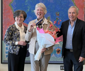
JoAnn Pushkin, Dr. Wendie Berg, and Dr Daniel Lehrer with Dr. Berg and her SBI Gold Medal on stage with flowers. Dr. Wendie Berg and her SBI Gold Medal on stage with flowers.


Receiving the SBI Gold Medal is both very gratifying and very humbling, and the words “gratifying” and “humbling” really sum up a career in breast imaging.  We are blessed by being able to truly make a difference in the lives of our patients every day, and mostly for the better.  I have been super fortunate to have had some impact on the field more broadly, and that gives me a deep sense of meaning.  But we all know that there is much still to be done.  I want to share some thoughts about a good friend of mine.  Susanne was a very successful research geneticist, very involved in her community, public health, and also in supporting political candidates who were interested in public health, communities, and scientific research.  She had dense breasts and knew to get screening MRI starting in 2007.  In 2009, the MRI showed what proved to be a 9-mm invasive lobular carcinoma.  Her nodes were negative and she had bilateral mastectomy.  She took aromatase inhibitors for ten years.  Still, despite all the favorable prognostic factors, in 2023, she was having abdominal discomfort, and was found to have carcinomatosis, and, soon thereafter, orbital metastases.  She signed up for many clinical trials and offered her tissue to researchers, recognizing that she was unlikely to personally benefit, but she felt it might help others in the future.  Sadly, Susanne passed almost exactly a year ago.  We have so much more to learn, and that sentiment is our shared legacy.

Today is an opportunity for me to publicly thank the research organizations, including NIH, the former Avon Foundation, the Breast Cancer Research Foundation, the Pennsylvania Breast Cancer Coalition, Komen Foundation, the Shear Family Foundation, and the many companies who have supported my research and other efforts.  I particularly want to thank the many, many national and international individuals who have helped me and supported me in this endeavor, and you know who you are, but especially Etta Pisano, Ellen Mendelson, Phil Evans, Daniel Lehrer, the many other investigators in ACRIN 6666 (which I just heard referred to as the “demon study”), and those behind the scenes (including Carl D’Orsi and the late Steve Feig).  Thank you to all those involved in DenseBreast-info.org, especially advocate and honorary SBI Fellow JoAnn Pushkin, and to the investigators in the trials comparing positron emission mammography and MRI, screening ultrasound and tomosynthesis, ultrasound AI, and my colleagues at the University of Pittsburgh now helping in studies of CEM.  I especially want to recognize the many women who have given their time and trust to participate in the many research studies I have led in the hopes that they will help others in the future. And today, I can say with confidence, that they have.  

Finally, I want to close with a bit more about Susanne.  She was a devoted and accomplished gardener, and one of her favorite sayings was “Friends are the flowers in the garden of life.”  I am blessed to have had many blooms in a rich garden of friends at the SBI.  We have accomplished so much together, and we have much more growing to do.  Thank you all.

– Dr. Wendie Berg, DenseBreast-info.org Chief Scientific Advisor

Filed Under: Blog

Supplemental Screening & the Latest Research

April 12, 2026 by JoAnn Pushkin

New Survey: Supplemental Screening ABUS Better Tolerated than MRI or CEM

DBI European Educator for Albania, and attending radiologist at the University of Cambridge and Cambridge University Hospitals, Dr. Iris Allajbeu, and colleagues, published a survey of 159 BRAID trial participants assessing patient acceptance, experience, and feedback for different methods of supplemental screening in women with dense breasts and negative mammography. Each of automated breast ultrasound (ABUS), abbreviated MRI (AB-MRI) and contrast-enhanced mammography (CEM) were generally well accepted by patients.


  • When compared to standard mammography, nearly all participants rated ABUS as the same or better; nearly 1 in 6 women rated AB-MRI as worse and 1 in 10 rated CEM as worse.
  • Mild or severe discomfort was common with ABUS and CEM.1
  • Anxiety before or during the imaging was least common with ABUS.
  • AB-MRI was well-received, with several women highlighting benefits, such as reduced pain due to less breast compression and improved detection accuracy. However, AB-MRI was associated with a higher proportion of negative feedback, suggesting that for some patients, its benefits may be outweighed by procedural drawbacks.
  • Withdrawal from the study was due to adverse patient experience for 151 women overall and was much more common with contrast-enhanced methods than with ABUS,2 making ABUS the most well tolerated imaging method.

Previously, we summarized the greater cancer detection rates from CEM and AB-MRI compared to ABUS in the first round of screening in the BRAID trial.3  Cancer detection rates were similar at 7-9.5/1000 across all three modalities in the second round of screening.3  False alarm rates were higher for CEM and AB-MRI in the first round and comparable across modalities in the second round.4

1 40/44 (90.1%) of ABUS participants reported discomfort as did 38/48 (79.2%) of CEM participants, compared to only 14/38 (36.8%) of MRI participants.

2 Of 151 withdrawals due to adverse patient experience, 69 were in women having CEM, 66 in women having AB-MRI, and only 12 in women having ABUS.

3 Cancer detection rates were 15/1572 (9.5/1000) for ABUS+MG; 9/1051 (8.6/1000) for AB-MRI+MG; and 6/870 (7.0/1000) for CEM in the second round. (By comparison, cancer detection rates in the first round were 4.2/1000 for ABUS; 17.4/1000 for AB-MRI; and 19.2/1000 for CEM after a negative mammogram.)

4 Recall rates in the second round were 4.3% for ABUS; 3.5% for AB-MRI; and 4.7% for CEM.  In the first round, recall rates were 4.0% for ABUS; 9.7% for AB-MRI; and 9.7% for CEM.


Updated Overview: International Atomic Energy Agency Panel Endorses Molecular Breast Imaging

An IAEA international expert panel has provided an updated overview of MBI using dual head detectors and 99mTc-sestamibi as a complement to mammography, especially in women with dense breasts or inconclusive conventional imaging. They conclude that “it remains a valuable modality in breast imaging, offering high sensitivity for the detection of small, node negative disease and detection of large, advanced cancers that are undetected by mammographic screening. MBI is performed at a safe radiation dose, is well-tolerated by patients, and has exceedingly low risk of adverse events.”

The ACR 2021 position is that MBI is “usually not appropriate” for screening in dense breasts due to whole body radiation exposure, though the radiation dose has decreased and additional promising prospective multicenter results have come out since that time.

Filed Under: Blog

New State Insurance Laws, State/Federal Initiatives

March 26, 2026 by JoAnn Pushkin

New Insurance Laws

Alabama and Wisconsin are the latest U.S. states to enact insurance laws for expanded no-cost breast imaging coverage. Based on DBI legislative analysis, there are now 38 states  (plus DC) that have enacted laws for expanded coverage (Minnesota’s is for diagnostic imaging only). The laws vary from state to state, and some still include out-of-pocket costs. Visit DBI’s legislation MAP for details by state.

Efforts to expand insurance coverage for extra testing are active at state and federal levels. To learn more about how state insurance laws intersect with federal initiatives like the new HRSA guidelines and the Find It Early Act, read our new content on the legislation MAP page (located below the map).

Filed Under: Blog

Resource Reminder + News from UK and Ireland

March 13, 2026 by JoAnn Pushkin

Screening Decision Support Flowchart
Resource reminder: Our popular decision support tool Who Needs More Screening? details evidence-based screening recommendations designed to optimize cancer detection in individuals in different risk categories, including dense breasts.


decision making flow chart cropped


UK News
Cheryl Cruwys, DBI’s European Education Coordinator and founder of Breast Density Matters UK, continues to lead organized lobbying efforts to the UK government. In a letter signed by 66 MPs to the Secretary of State for Health and Social Care, urgent action was requested for “density inform” measures and needed policy changes.

6 UK MPs in front of Breast Density Matters UK banner


Ireland News
The Health Information and Quality Authority has published a protocol to assess potential modifications to the BreastCheck program to consider ways in which breast density could be incorporated into the mammography screening pathway.

Filed Under: Blog

Dense Breast Progress on Two Fronts: New Trial Results and State Bills Head to Governors

March 3, 2026 by JoAnn Pushkin

State Insurance Bills

Bills in both Alabama and Wisconsin have advanced to the Governor’s desk. Stay tuned for more details if/when the bills are signed into law.

J-START TRIAL FOLLOW-UP

The J-START, a prospective Japanese randomized trial that compared Mammography plus Ultrasound (US, intervention group) to Mammography alone (control group) in women age 40 to 49 years of age previously showed, as primary endpoints, that the intervention of adding US twice in two years improved detection of early stage (0 or 1) breast cancer and reduced interval cancers, but lowered specificity. In a subset analysis, US improved early breast cancer detection in both nondense and dense breasts.

Longterm follow-up (median exceeding 11 years) is now available, showing significant reduction in advanced stage cancers (stage II or higher: node positive or T2 or both) in the intervention group.

  • 234/894 (26%) carcinomas were advanced stage at diagnosis in the intervention group
  • 277/843 (33%) carcinomas were advanced in the control group.
  • Hazard ratio for advanced stage at diagnosis was 0·83, 95·6% CI 0·70–0·98; p=0·026)
  • Divergence between groups emerged around year four, widened until year eight, and remained stable thereafter.

In an accompanying editorial, DBI Medical Advisory Board members Drs Jean Seely and Paula Gordon point out that the magnitude of reduction in advanced cancers due to addition of US in women 40-49 is similar to the magnitude of mortality reduction attributable to screening mammography in that age group across randomized trials. These results are quite compelling for Asian women, though may not generalize to all populations. In many other studies in other patient populations, US has been shown to substantially improve early breast cancer detection when added to annual or biennial 2D mammography in women with dense breasts. The benefits of screening US are very small, however, in women having annual tomosynthesis.

Filed Under: Blog

We’re Reaching Patients and Providers—Everywhere

February 23, 2026 by JoAnn Pushkin

Multi-lingual Patient Flyers

Our patient flyer, Why a Conversation About Your Breast Density Matters is available in over 30 languages and dialects. The one-page printable flyer is an ideal resource for clinics or health fairs. Share confidently, all DBI translated content is meticulously crafted by native speakers specializing in breast radiology.


Lecture: When is Screening Mammography Not Enough?

Dr. Wendie Berg and Dr. Philip Strax

DBI’s Chief Scientific Advisor, Dr. Wendie Berg, was honored to give the Philip Strax, MD Breast Imaging and Saving Lives Lecture for the Texas Radiological Society in Houston on February 14th, entitled “When is Screening Mammography Not Enough?” Dr. Strax is the radiologist who, together with statistician Sam Shapiro, led the pivotal and pioneering Health Insurance Plan of New York randomized trial of screening mammography in the 1960s.  Dr. Richard Strax, also a radiologist, has honored his father’s legacy with this lectureship. Dr. Daniel Kopans, Dr. Edward Sickles, and Dr. Debra Monticciolo are previous presenters.

Highlights from Dr. Berg’s presentation included:

  • Participation in screening is the biggest issue needing improvement, including identifying women at higher risk who should start screening earlier than age 40 (and usually with MRI).  To accomplish this, all women ideally should have risk assessment by age 25, especially those of Black, Hispanic, Asian, or Ashkenazi Jewish descent or with family history of breast or ovarian cancer.
  • Breast density, methods to assess risk, and supplemental screening options, with emphasis on when MRI is indicated, and gaps in current insurance coverage that would be addressed by the Find It Early Act.
  • The “flip side” of risk assessment of possibly reducing screening of women based on low risk, including the WISDOM trial results. Such approaches are limited by concerns that current methods (including AI) are inadequate to identify all women who will develop cancer in the next 2-5 years.
  • A look to the future may include liquid biopsy as a screening method, with the potential that breast imaging would become more focused on diagnostic imaging and biopsy


Newly Appointed

DBI’s European Educator and supporter for France, Professor Isabelle Thomassin-Naggara, MD, has been appointed as ESR Quality, Safety & Standards Committee (QSSC) Chair.  This is another distinguished appointment for Prof. Thomassin-Naggara, who also holds the post as Women’s Imaging Editor at the European Journal of Radiology. We extend our congratulations to, Isabelle!

Filed Under: Blog

Legislation + Education Updates in the U.S. and Abroad

February 16, 2026 by JoAnn Pushkin

Find It Early Act – Updated Statement of Support

DBI has played a pivotal role in drafting and advocating for this landmark legislation. The bill aims to eliminate patient costs for supplemental and diagnostic breast imaging for women with dense breasts or at increased risk, and close gaps in current state laws and federal guidelines. Crucially, it extends coverage across private and public plans, including Medicare, TRICARE, and the VHA. DBI has coordinated the latest update of the FIEA Statement of Support, now backed by a coalition of 50 leading organizations. To contact your federal representative to ask for their support of this bill, please visit FindItEarlyAct.org for lobbying tools, the Statement of Support, and FIEA Fact Sheet .

Canadian Society of Breast Imaging – Position Statement

The Canadian Society of Breast Imaging published their updated position statement on mammographic breast density and supplemental screening in the Journal of Breast Imaging.  The statement, co-authored by DBI Medical Advisory Board members Paula Gordon, MD and Jean Seely MD, includes the following key recommendations for Canadian women:

  • All women should begin regular screening mammography with digital breast tomosynthesis (DBT) (or standard 2D mammography if DBT is unavailable) every 1-2 years beginning at age 40, unless individual risk assessment indicates earlier screening.
  • Because vascular-based imaging (MRI or CEM) offers the highest sensitivity and specificity, women at average risk with heterogeneously or extremely dense breasts (ACR density categories C or D) should be offered supplemental screening in the following order of preference: MRI every 2 years, contrast-enhanced mammography every 2 years, or ultrasound annually.
  • If availability of supplemental screening is limited, access should be prioritized for women with extremely dense breasts (ACR density category D).
  • Women considered high risk for breast cancer (lifetime risk 20%-25% or greater) should have both annual mammography beginning at age 30 years and annual MRI screening from ages 25-69 years. Mammography alone is recommended to continue at age 70 and beyond.
Europe / UK Education

At the invitation of the University of Exeter, Cheryl Cruwys—DBI’s European Education Coordinator—will deliver a lecture to diagnostic radiography students on February 17th. Her presentation will cover the complexities of breast density and provide an overview of the professional healthcare resources available on the DBI website.

Filed Under: Blog

Hawaii and Utah Add Bills + MASAI Trial

February 10, 2026 by JoAnn Pushkin

New State Insurance Bills

Two new state bills for expanded coverage for breast imaging: Hawaii HB2366, Utah HB0468. For existing state insurance laws, see our MAP.

color coded map of the united states

MASAI Trial

A large, randomized, prospective, controlled, single-blinded non-inferiority trial including nearly 106,000 women in the Swedish breast cancer screening program was conducted to compare interval cancer rates between women screened with standard double reading versus Transpara AI-supported mammography to triage mammograms that should undergo double reading. The study included a mix of tomosynthesis and 2D mammography. Interval cancers were defined as primary breast cancers diagnosed between two screening rounds (typically every 1.5 – 2 years in average-risk women) or within 2 years after the last scheduled screening that were not detected at screening.

  • The interval cancer rate was lower among women who received AI-supported mammography compared to those who had standard double reading.1
  • This effect was consistent across age and breast density.
  • Sensitivity was higher in the AI-supported mammography group2 and specificities were equal among both groups.3

AI-supported mammography improved cancer detection, reduced interval cancer rates and reduced radiologist workload by decreasing the need for double reading.  In the United States, double reading is not routinely performed and isolated AI interpretation is not yet accepted: there are barriers to implementation. Further, AI is not generally covered by insurance so facilities in the USA may opt to charge patients an out-of-pocket fee when offering AI.

1 Interval cancer rates were 1.55/1000 (82/53043) (95% CI 1·23–1·92) and 1·76/1000 (93/52872) (95% CI 1·42–2·15) in the intervention and control groups respectively, with a non-inferior proportion ratio of 0·88 (95% CI 0·65–1·18; p=0·41).

2 Sensitivities were 80.5% (338/420) and 73.8% (262/355) in the AI-supported and standard double reading groups, respectively.

3 Specificity was 98.5% in both groups (51852/52623 in the AI supported group vs. 51752/52517 in the double reading group).

Filed Under: Blog

State Legislative Initiatives Continue + New Research

January 31, 2026 by JoAnn Pushkin

New State Insurance Bills

State legislative initiatives for expanded coverage for breast imaging continue. This week’s bill introductions:  Kansas SB409, New Jersey A1462, New Jersey A1945, New Jersey A2152, New Jersey A2775.

For details on existing state insurance laws, visit the DenseBreast-info.org MAP.


New Study

A large study led by Ariel S. Kniss, MD, PhD evaluated the impact of breast density on performance of screening mammography with combination digital 2D mammography and tomosynthesis (DBT) from over 300,000 digital breast tomosynthesis (DBT) exams from over 111,000 women. All screening DBT exams were performed from January 2013 to June 2019 at one institution. Density was categorized using visual BI-RADS categories (A, almost entirely fatty, B, scattered areas of fibroglandular density, C, heterogeneous dense tissue, or D extremely dense tissue).

  • Sensitivity of screening DBT decreased with increasing density1
  • Specificity of screening DBT was lower in women with category D density than those with category A, B, or C, respectively. 2

Performance of screening DBT decreases with increasing breast density, reinforcing the importance of supplemental screening considerations in women with dense breasts, even when DBT is used for screening mammography.

1 Sensitivities were 92.8%, 90.1%, 81.0%, and 61.8% for categories A, B, C, and D, respectively (P < 0.001 for category D vs. A, B, or C, respectively).

2 Specificities were 96.7%, 94.4%, 92.5%, and 93.3% for categories A, B, C, and D (P < 0.001 for category D vs. A).

Filed Under: Blog

  • « Go to Previous Page
  • Page 1
  • Page 2
  • Page 3
  • Page 4
  • Interim pages omitted …
  • Page 22
  • Go to Next Page »

Primary Sidebar

DenseBreast‑info, Inc. is an independent, nonprofit organization. DBI is not affiliated with or endorsed by other organizations unless expressly identified. If you have questions about DBI’s programs or partnerships, please contact us.

© Unpublished, DenseBreast-info, Inc. ALL RIGHTS RESERVED | 501(c)(3) Public Charity

  • Online Privacy Policy
  • Online Terms of Use & Disclaimers

This website is for informational purposes only and is not intended to be a substitute for medical advice from a physician or to create a standard of care for health care providers. In addition, information has been provided by independent third parties and may not represent the views of DenseBreast-info.org. Please check with a physician regarding your specific condition, if you have any questions, or need a diagnosis. If you are experiencing an urgent medical condition, call your local emergency services [e.g., 9-1-1 (in the USA) or 1-1-2 (in Europe)].